First PSMA PET/MRI Scan in georgia
By Amol Takalkar*
The first prostate specific membrane antigen (PSMA) radiotracer positron emission tomography/magnetic resonance imaging (PET/MRI) scan in Georgia was recently performed on the new GE Signa PET/MRI at Winship Cancer Institute at the Emory Clinic by the Department of Radiology and Imaging Sciences.
The PSMA PET/MRI (depicted in the figure below) was performed in a patient with biochemical relapse of prostate cancer. He was diagnosed with high-risk prostate cancer and had been treated with radiation therapy and hormonal therapy. Over the past year, his PSA levels rose from undetectable to 2.16 ng/mL indicating biochemical recurrence of prostate cancer.
The PSMA PET/MRI (depicted in the figure below) was performed in a patient with biochemical relapse of prostate cancer. He was diagnosed with high-risk prostate cancer and had been treated with radiation therapy and hormonal therapy. Over the past year, his PSA levels rose from undetectable to 2.16 ng/mL indicating biochemical recurrence of prostate cancer.
PSMA PET/MRI scan
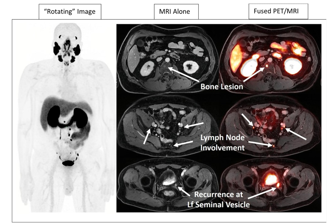
The figure above shows a “rotating” image on the left and on the right, selected slices of the MRI and fused PET/MRI showing bone and lymph node prostate cancer metastasis, as well as local recurrence in the left seminal vesicle. The nodes were small and would have been considered normal on MRI alone and even the bone lesion was also difficult to see on MRI alone. The lesions were strikingly intense due to uptake of the PSMA radiotracer on PET depicting the high contrast of this specific radiotracer for prostate cancer lesions.
18F-DCFPyL (F-18 piflufolastat, trade name: Pylarify) is a fluorinated small molecule PSMA PET probe specifically targeting PSMA, a protein expressed in high concentrations in most prostate cancer cells. This radiotracer was FDA approved in May 2021 for PET imaging of high-risk primary and recurrent prostate cancer.
PET/MRI is a novel hybrid imaging technology combining the anatomic and tissue characterization strengths of MRI with the functional imaging capabilities of PET to provide exquisite images with excellent tumor-to- background contrast and exceptional tissue characterization. This scanner was installed at Emory in 2021 and is one in a small number of institutions in the US, and the only one in Georgia to offer this novel and highly specialized integrated imaging modality. It is expected to provide deeper insights into cancer, neurology, and cardiology diseases.
This exciting advancement is made possible by a highly skilled imaging team. Dr. Shahein Tajmir, clinical modality director for PET-MR, spearheaded operationalization of this service. The team also includes Michael Larche, PET-MR supervisor, and PET-MR imaging technologists Nichole Dickerson and Leah Mims.
PSMA targeted imaging which can be used with PET/CT or PET/MRI is expected to cause a paradigm shift in the current workup of prostate cancer patients with suspected metastases or recurrent disease and will lead to more accurate and early identification disease sites than conventional imaging. This will have a strong impact on treatment choices and decisions and hopefully better outcomes for patients. PSMA based PET imaging is also expected to revolutionize management of prostate cancer by paving the way for development and implementation of PSMA based therapy approaches for treating metastatic prostate cancer in the near future.
*Amol Takalkar, MD, MS, MBA, FACNM, is a professor of radiology & imaging sciences in the Division of Nuclear Medicine & Molecular Imaging of the Emory University School of Medicine’s Department of Radiology and Imaging Sciences